The Lateral Cervical Region
By : Karam AlanazDefinition
it is triangle in shape , that divided into a large occipital triangle superiorly and a small omoclavicular triangle (supraclavicular) inferiorly by the inferior belly of the omohyoid.
Boundaries
Anteriorly : the posterior border of the SCM(sternocleidomastoid).
Posteriorly : the anterior border of the trapezius.
Inferiorly : the middle third of the clavicle between the trapezius and the SCM.
And by an apex, where the SCM and trapezius meet on the superior nuchal line of the occipital bone.
Roof
formed by:
(1) skin .
(2) Superficial fascia that contains : platysma muscle , Cutaneous branches of Cervical plexus, External Jugular vein .
(3) deep fascia by investing layer .
Floor
formed by:
muscles covered by the prevertebral layer of deep cervical fascia.
Posteriorly : the anterior border of the trapezius.
Inferiorly : the middle third of the clavicle between the trapezius and the SCM.
And by an apex, where the SCM and trapezius meet on the superior nuchal line of the occipital bone.
Roof
formed by:
(1) skin .
(2) Superficial fascia that contains : platysma muscle , Cutaneous branches of Cervical plexus, External Jugular vein .
(3) deep fascia by investing layer .
Floor
formed by:
muscles covered by the prevertebral layer of deep cervical fascia.
NOTES
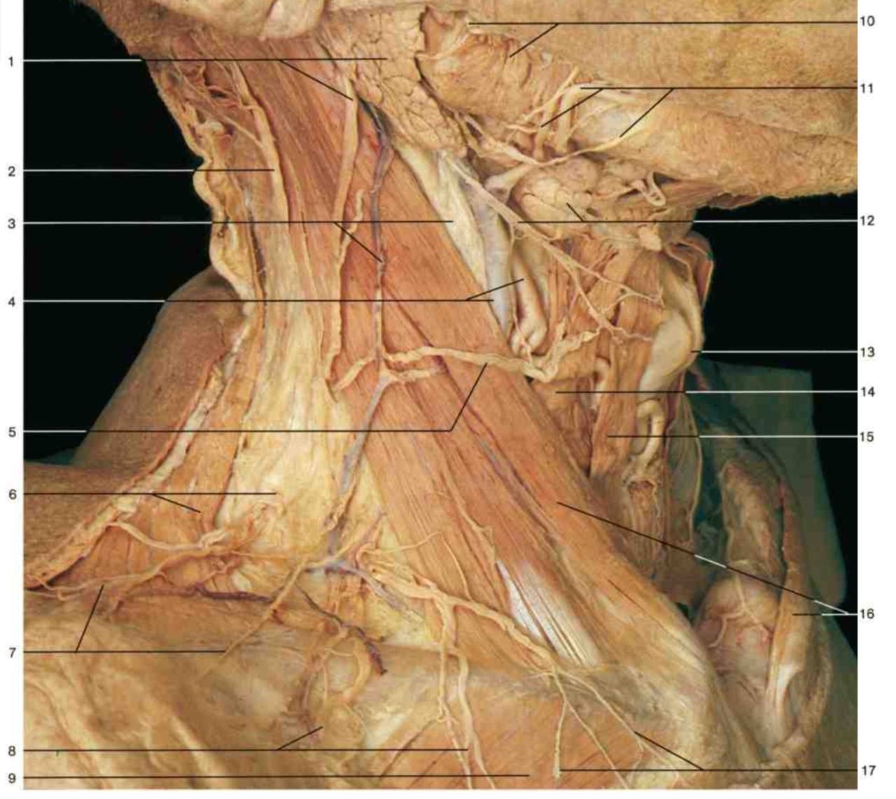
1-The floor of the posterior triangle is usually formed by the prevertebral fascia overlying four muscles : splenius capitis, levator scapulae, middle scalene, and posterior scalene. Sometimes the inferior part of the anterior scalene appears in the inferomedial angle of the triangle , where it is usually hidden by the SCM.
2-The occipital triangle is so called because the occipital artery appears in its apex . The most important nerve crossing the occipital triangle is the spinal accessory nerve (CN XI).
The omoclavicular triangle is indicated on the surface of the neck by the supraclavicular fossa.
1-The floor of the posterior triangle is usually formed by the prevertebral fascia overlying four muscles : splenius capitis, levator scapulae, middle scalene, and posterior scalene. Sometimes the inferior part of the anterior scalene appears in the inferomedial angle of the triangle , where it is usually hidden by the SCM.
2-The occipital triangle is so called because the occipital artery appears in its apex . The most important nerve crossing the occipital triangle is the spinal accessory nerve (CN XI).
The omoclavicular triangle is indicated on the surface of the neck by the supraclavicular fossa.
Content
CONTENTS OF THE POSTERIOR TRIANGLE :
1- MUSCLE
inferior belly of omohyoid , it is a slender muscle which arises from the upper border of scapula & supra-scapular ligament .
2- ARTERIES OF POSTERIOR TRIANGLE
(A) 3rd part of subclavian artery : lies in the lower part of the triangle Close to the lower trunk of the brachial plexus it extends from the lateral border of scalenus anterior to the outer border of 1st rib where it continues as axillary artery.
(B)Suprascapular artery : it is a branch of thyrocervical trunk of 1st part of subclavian artery it reaches the posterior triangle by crossing infront of scalenus anterior muscle then descends behind the Clavicle (with its nerve) to share in the anastomosis around the scapula
(C) transverse Cervical artery : it is another branch of thyrocervical trunk which passes above the suprascapular a to reach the posterior triangle, where it ends on levator scapulae by dividing into:
(a) superficial branch (b) deep branch
(D) 3rd part of occipital artery (branch of external carotid artery) :crosses the apex of the posterior triangular then disappears deep to the trapezius muscle .
3- VEINS OF POSTERIOR TRIANGLE
(1)Subclavian Vein : it lies in the lower most part of the posterior triangle mostly behind the clavicle.
(2) External Jugular Vein : it descends in the superficial fascia to a point which above the clavicle where it pierces the deep fascia of the roof of posterior triangle .
4- NERVES OF POSTERIOR TRIANGLE
(1) Spinal accessory nerve : it appears at the middle of posterior-border of sternomastoid , it crosses the posterior triangle over levator scapulae being adherent to the fascial roof of the triangle , it disappears deep to the anterior -border of trapezius muscle -about 5 cm above the clavicle. it supplies both stemomastoid & trapezius muscles .
(2) Brachial plexus : only the roots & the trunks of the plexus appear in the posterior triangle between the scalenus anterior & scalenus medius muscle above & behind the subclavian artery the upper trunk gives 2 branches in the posterior triangle:
(a) the suprascapular nerve (b)nerve-to subclavius muscle
(3) Cervical plexus and its Cutaneous branches appear in the posterior triangle at the middle of posterior-border of sternomastoid muscle.
1- MUSCLE
inferior belly of omohyoid , it is a slender muscle which arises from the upper border of scapula & supra-scapular ligament .
2- ARTERIES OF POSTERIOR TRIANGLE
(A) 3rd part of subclavian artery : lies in the lower part of the triangle Close to the lower trunk of the brachial plexus it extends from the lateral border of scalenus anterior to the outer border of 1st rib where it continues as axillary artery.
(B)Suprascapular artery : it is a branch of thyrocervical trunk of 1st part of subclavian artery it reaches the posterior triangle by crossing infront of scalenus anterior muscle then descends behind the Clavicle (with its nerve) to share in the anastomosis around the scapula
(C) transverse Cervical artery : it is another branch of thyrocervical trunk which passes above the suprascapular a to reach the posterior triangle, where it ends on levator scapulae by dividing into:
(a) superficial branch (b) deep branch
(D) 3rd part of occipital artery (branch of external carotid artery) :crosses the apex of the posterior triangular then disappears deep to the trapezius muscle .
3- VEINS OF POSTERIOR TRIANGLE
(1)Subclavian Vein : it lies in the lower most part of the posterior triangle mostly behind the clavicle.
(2) External Jugular Vein : it descends in the superficial fascia to a point which above the clavicle where it pierces the deep fascia of the roof of posterior triangle .
4- NERVES OF POSTERIOR TRIANGLE
(1) Spinal accessory nerve : it appears at the middle of posterior-border of sternomastoid , it crosses the posterior triangle over levator scapulae being adherent to the fascial roof of the triangle , it disappears deep to the anterior -border of trapezius muscle -about 5 cm above the clavicle. it supplies both stemomastoid & trapezius muscles .
(2) Brachial plexus : only the roots & the trunks of the plexus appear in the posterior triangle between the scalenus anterior & scalenus medius muscle above & behind the subclavian artery the upper trunk gives 2 branches in the posterior triangle:
(a) the suprascapular nerve (b)nerve-to subclavius muscle
(3) Cervical plexus and its Cutaneous branches appear in the posterior triangle at the middle of posterior-border of sternomastoid muscle.

Superficial Veiw of Post.triangle
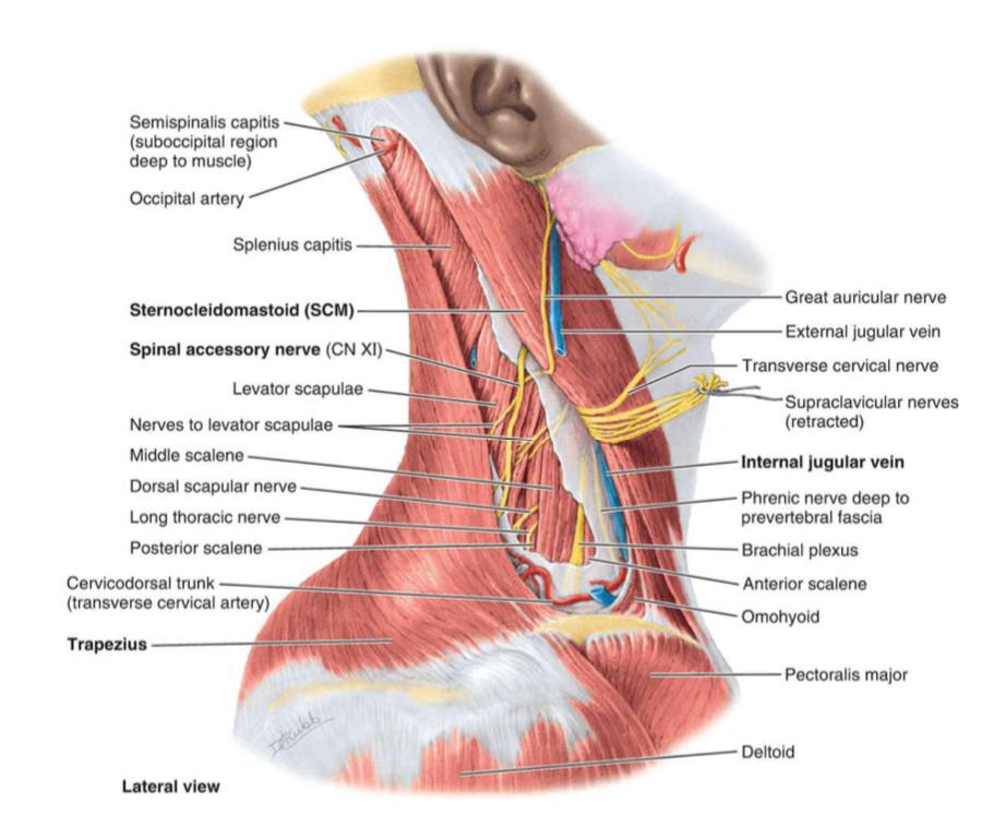
Cutaneous Branches of cervical plexus
1) Lesser occipital nerve: it arise from anterior ramus of (C2) , it supplies the skin of the upper 2 of inner surface of the auricle and the adjoining part of the scalp.
(2) the Great auricular nerve :- it arise from anterior ramus of (C2,C3), it supplies the skin over the angle of mandible , the skin over the parotid gland and the skin of the lower of the outer & inner surfaces of the auricle.
(3) the transverse (anterior) cutaneous nerve of neck : it arise from (C2,3) , it divides into upper & lower branches supplying the skin of the anterior .
(4)the supraclavicular nerve: it arise from (C3,C4), it divides into 3 branches (medial, intermediate & lateral) which descend superficial to the clavicle supplying: skin of the posterior triangle of neck , skin over tip of shoulder and skin of Chest as far down as the sternal angle (2nd rib)
(2) the Great auricular nerve :- it arise from anterior ramus of (C2,C3), it supplies the skin over the angle of mandible , the skin over the parotid gland and the skin of the lower of the outer & inner surfaces of the auricle.
(3) the transverse (anterior) cutaneous nerve of neck : it arise from (C2,3) , it divides into upper & lower branches supplying the skin of the anterior .
(4)the supraclavicular nerve: it arise from (C3,C4), it divides into 3 branches (medial, intermediate & lateral) which descend superficial to the clavicle supplying: skin of the posterior triangle of neck , skin over tip of shoulder and skin of Chest as far down as the sternal angle (2nd rib)
lymph nodes
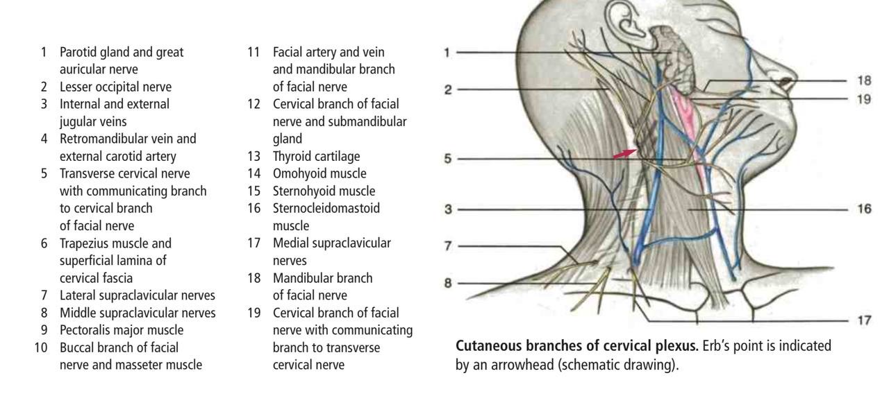
1) Occipital Lymph node : at the apex of the triangle alongside the occipital artery.
(2) Supraclavicular Lymph node :at the base of the triangle on the subclavian artery.
(2) Supraclavicular Lymph node :at the base of the triangle on the subclavian artery.
Referance
1-Anatomy of upper limb by Sameh Doss , p.151, p152,p153
2- Moore-clinically oriented-Anatomy eighth edition , p.2237, p.2238
Figures:
Moore-clinically oriented-Anatomy eighth edition ، figure 9.9 superficial dissection of lateral cervical region .
Moore-clinically oriented-Anatomy eighth edition, Figure 9.10 deep dissection of lateral cervical region.
color Atlas of anatomy seventh edition,p.178,p179,p.180-Regions of Neck - lateral region
Atlas of human anatomy sixth edition - nerves and vessels of the neck, p.50
2- Moore-clinically oriented-Anatomy eighth edition , p.2237, p.2238
Figures:
Moore-clinically oriented-Anatomy eighth edition ، figure 9.9 superficial dissection of lateral cervical region .
Moore-clinically oriented-Anatomy eighth edition, Figure 9.10 deep dissection of lateral cervical region.
color Atlas of anatomy seventh edition,p.178,p179,p.180-Regions of Neck - lateral region
Atlas of human anatomy sixth edition - nerves and vessels of the neck, p.50