Sacrospinous ligament
By : Banan AlzubaidiDefinition :
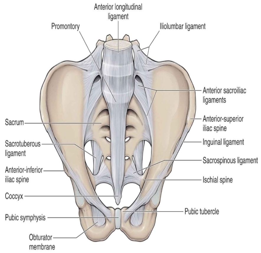
. Sacrospinous ligament : it is a thin strong triangular band (from
connective tissue)(it is considered anterior and small sacrosciatic
ligament ).
. about (3.8-4.6) in length .
. it is a stabilizer of the sacroiliac
joint and connects the pelvic bone to the vertebral column.
. It is converts
the sciatic notches to the sciatic foramina .. and forms a part of the
sciatic foramina border.
connective tissue)(it is considered anterior and small sacrosciatic
ligament ).
. about (3.8-4.6) in length .
. it is a stabilizer of the sacroiliac
joint and connects the pelvic bone to the vertebral column.
. It is converts
the sciatic notches to the sciatic foramina .. and forms a part of the
sciatic foramina border.
Function:
. sacrospinous ligament and sacrotuberous ligament assist
in the stability of the pelvic bone and work together to prevent
rotation of the ilium past the sacrum and prevent excessive twisting
of the pelvis and limit downhill of the sacrum.
in the stability of the pelvic bone and work together to prevent
rotation of the ilium past the sacrum and prevent excessive twisting
of the pelvis and limit downhill of the sacrum.
Origin and insertion :
. origin: anterolateral of the sacrum(s2-s3-s4)and coccyx(tail bone).
. insertion : ischial spine (part of the pelvic bone)
. insertion : ischial spine (part of the pelvic bone)
Relations :
. anteriorly : coccygeus muscle (extends from the ischial spine (part of the pelvic
bone )to the inferior end of the sacrum and coccyx).
. posteriorly: sacrotuberous ligament .
. superiorly: upper margin of the ligament forms the lower border of the greater
sciatic foramen.
. inferiorly: lower margin of the ligament forms the upper border of the lesser
sciatic foramen
bone )to the inferior end of the sacrum and coccyx).
. posteriorly: sacrotuberous ligament .
. superiorly: upper margin of the ligament forms the lower border of the greater
sciatic foramen.
. inferiorly: lower margin of the ligament forms the upper border of the lesser
sciatic foramen

NOTES :
. the sacrospinous ligaments fibers overlap with the fibers of the sacrotuberous
ligament.
. the sacrospinous and sacrotuberous ligaments are most stressed when the
person leans forward or stands up.
. The sacrospinous ligament are surrounded by the pudendal nerve and
vessels(artery and vein), the sciatic nerve and the inferior gluteal artery .
ligament.
. the sacrospinous and sacrotuberous ligaments are most stressed when the
person leans forward or stands up.
. The sacrospinous ligament are surrounded by the pudendal nerve and
vessels(artery and vein), the sciatic nerve and the inferior gluteal artery .

Clinical note :
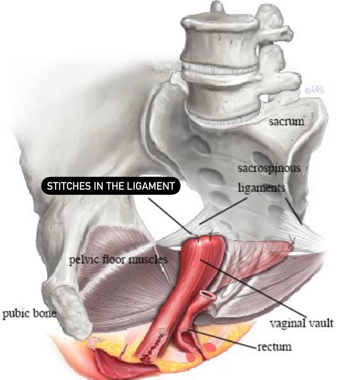
. sacrospinous ligament suspension : It is a surgical procedure to restore
support to the upper part of the vagina after a prolapse of the uterus
and vagina.
. It often occurs after a hysterectomy and can be
performed with the uterus remaining in place.
. The operation is
performed extraperitoneally, meaning that the surgeon does not
enter the pelvic cavity.
. This surgical intervention involves stitching the sacrospinous
ligament to the vaginal vault or cervix in order to restore support to
treat that case.
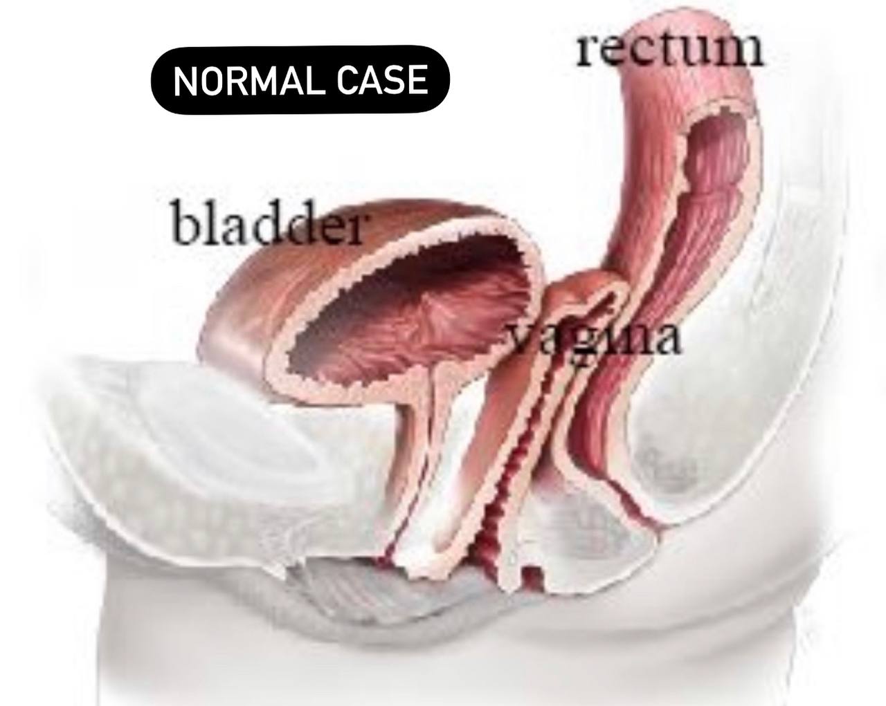
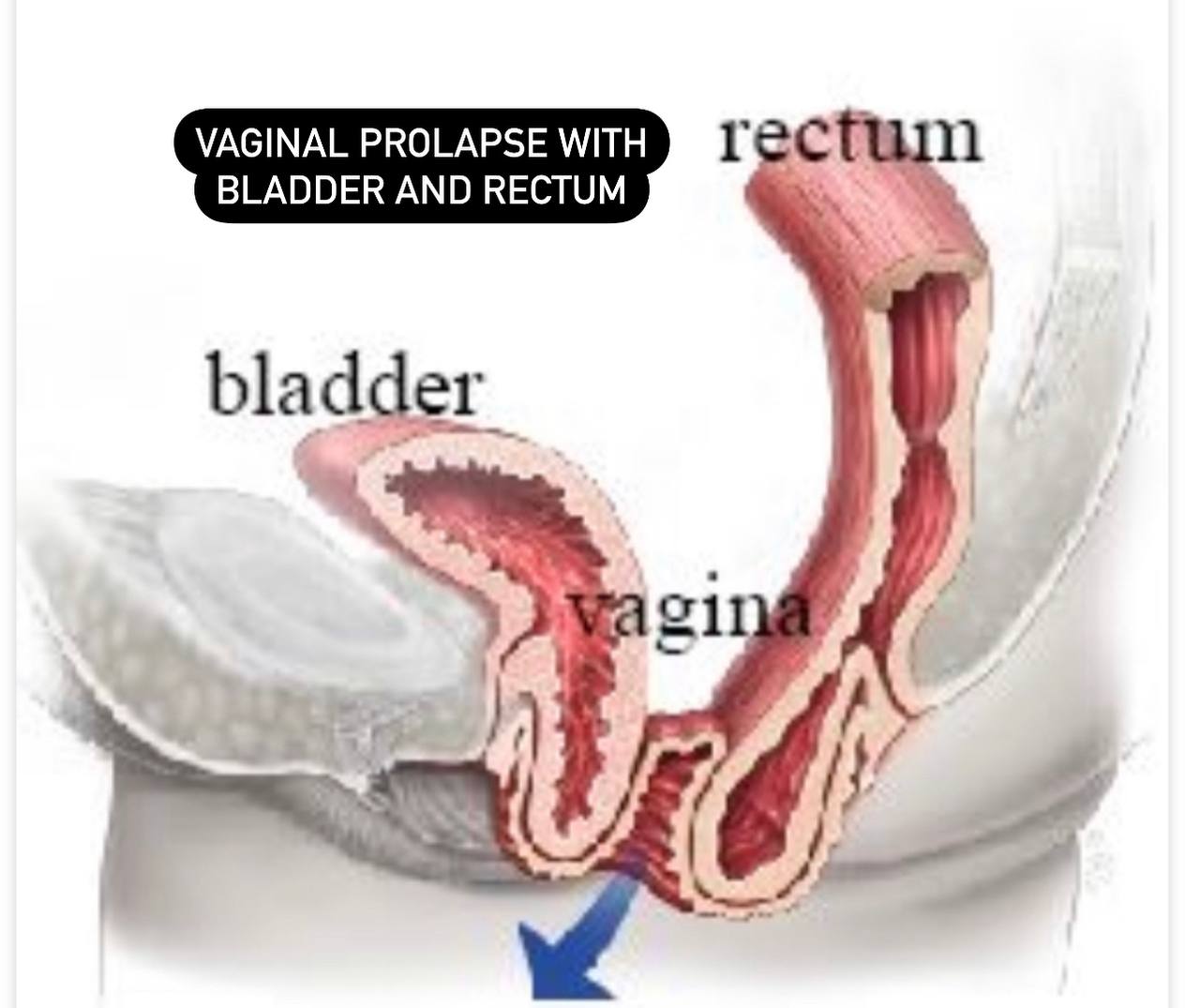
. Prolapse of the vagina or pelvic organs : it occurs when the
ligaments in the pelvic and surrounding tissues become damaged or
due to pelvic floor weakness due to childbirth or postmenopausal
atrophy, and vaginal prolapse may occur after hysterectomy....or as a
result of a damage to the supporting structures of the uterus and
vagina this cause prolapse or falling of the vagina or uterus.
support to the upper part of the vagina after a prolapse of the uterus
and vagina.
. It often occurs after a hysterectomy and can be
performed with the uterus remaining in place.
. The operation is
performed extraperitoneally, meaning that the surgeon does not
enter the pelvic cavity.
. This surgical intervention involves stitching the sacrospinous
ligament to the vaginal vault or cervix in order to restore support to
treat that case.
. Prolapse of the vagina or pelvic organs : it occurs when the
ligaments in the pelvic and surrounding tissues become damaged or
due to pelvic floor weakness due to childbirth or postmenopausal
atrophy, and vaginal prolapse may occur after hysterectomy....or as a
result of a damage to the supporting structures of the uterus and
vagina this cause prolapse or falling of the vagina or uterus.
Symptoms of prolapse of the vagina or pelvic organs:
1) Mixed urinary incontinence.
2) Colorectal symptoms (constipation and indigestion) .
3) Anal symptoms such as fecal incontinence.
4) Swelling in the vagina and a feeling of fullness and heaviness in the vagina or an
external bulge which extends outside the vagina.
5) Difficulty urinating and bowel movement.
6) Difficulty in intercourse.
2) Colorectal symptoms (constipation and indigestion) .
3) Anal symptoms such as fecal incontinence.
4) Swelling in the vagina and a feeling of fullness and heaviness in the vagina or an
external bulge which extends outside the vagina.
5) Difficulty urinating and bowel movement.
6) Difficulty in intercourse.
This process is done through the following steps:
. During this surgery, patients are placed on their back with their legs
in support stirrups.
. This surgery is done through an incision in the vagina.
. The ligament is identified and removed from the surrounding tissue.
. Permanent or slowly dissolving stitches are placed in the
sacrospinous ligament( It is a formed from the ligament with the
coccyx muscle (a main muscle)..so the ligament provides a key point
for fixing the top of the vagina).
. The ligament is suspended with the pubis. Then through the upper
part of the vagina, tissues that support the vagina are formed.
in support stirrups.
. This surgery is done through an incision in the vagina.
. The ligament is identified and removed from the surrounding tissue.
. Permanent or slowly dissolving stitches are placed in the
sacrospinous ligament( It is a formed from the ligament with the
coccyx muscle (a main muscle)..so the ligament provides a key point
for fixing the top of the vagina).
. The ligament is suspended with the pubis. Then through the upper
part of the vagina, tissues that support the vagina are formed.
Complications of this operation:
. Usually, the complications are very few during this operation ,but
with every operation there are risks:
1) Pain in the buttocks during the first weeks of the operation (the
most common).
2) Possible bleeding and the formation of a hematoma (very rare).
3) Or it may occur Vaginitis, bladder or urinary tract infection (rare)
with every operation there are risks:
1) Pain in the buttocks during the first weeks of the operation (the
most common).
2) Possible bleeding and the formation of a hematoma (very rare).
3) Or it may occur Vaginitis, bladder or urinary tract infection (rare)
NOTES :
. This procedure is often combined with a vaginal hysterectomy or surgery to treat
the prolapsed bladder, bowel or rectum and urinary incontinence.
. Additional prolapse procedures such as anterior colitis and posterior colitis are
done to correct any additional prolapse of the vaginal walls.
the prolapsed bladder, bowel or rectum and urinary incontinence.
. Additional prolapse procedures such as anterior colitis and posterior colitis are
done to correct any additional prolapse of the vaginal walls.
Other clinical note :
. The pudendal nerve may become involved between the sacrotuberous
ligament and the sacrospinous ligament (this the inner space of these
ligaments).
. This causes chronic perineal pain ..the sacrotuberous ligamemt is a
major factor in the undiagnosed chronic perineal pain.
ligament and the sacrospinous ligament (this the inner space of these
ligaments).
. This causes chronic perineal pain ..the sacrotuberous ligamemt is a
major factor in the undiagnosed chronic perineal pain.
References of images :
Cover image/sacrospinous ligament /Anatomy zone /https://anatomyzone.com/articles/sacrospinous-ligament/
Fig. 1/SNELLŚ CLINICAL ANATOMY BY REGIONS/Figure 10.8 / Course and branches of the pudendal nerve in the
male.
Fig. 2/sacrospinous ligament //musculoskeletalkey/https://musculoskeletalkey.com/the-sacroiliac-joint-2/
Fig.3/sacrospinous ligament /Wiley online library /https://onlinelibrary.wiley.com/doi/10.1002/ca.23328
Fig. 4/sacrospinous Fixation/Your pelvic floor/https://www.yourpelvicfloor.org/conditions/sacrospinous-fixation/
Fig. 5/sacrospinous fixation_IIIeococcygeus suspension/Your pelvic floor/https://www.yourpelvicfloor.org/conditions/sacrospinous-fixation/
Fig. 6/sacrospinous ligament _IIIeococcygeus suspension/Your pelvic floor/https://www.yourpelvicfloor.org/conditions/sacrospinous-fixation/
Fig. 1/SNELLŚ CLINICAL ANATOMY BY REGIONS/Figure 10.8 / Course and branches of the pudendal nerve in the
male.
Fig. 2/sacrospinous ligament //musculoskeletalkey/https://musculoskeletalkey.com/the-sacroiliac-joint-2/
Fig.3/sacrospinous ligament /Wiley online library /https://onlinelibrary.wiley.com/doi/10.1002/ca.23328
Fig. 4/sacrospinous Fixation/Your pelvic floor/https://www.yourpelvicfloor.org/conditions/sacrospinous-fixation/
Fig. 5/sacrospinous fixation_IIIeococcygeus suspension/Your pelvic floor/https://www.yourpelvicfloor.org/conditions/sacrospinous-fixation/
Fig. 6/sacrospinous ligament _IIIeococcygeus suspension/Your pelvic floor/https://www.yourpelvicfloor.org/conditions/sacrospinous-fixation/
Reference of the video :
Sacrospinous ligament Fixation (sacrospinous hysteropexy) /geogyn1/https://youtu.be/ySSfy2A1_RM